
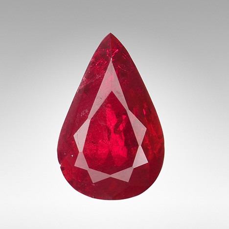
The Tokyo laboratory recently received a 3.02 ct red pear-shaped mixed cut measuring 10.93 × 7.04 × 5.03 mm (figure 1). Standard gemological testing yielded a refractive index of 1.765–1.773; a hydrostatic specific gravity of 4.00; absorption lines at 693, 476, and 468 nm; and a broad absorption band between 500 nm and 610 nm seen using a handheld spectroscope. All of these properties were consistent with ruby. The stone displayed strong red fluorescence under both long-wave and short-wave UV.
Figure 2. These natural-looking hexagonal colorless crystals and the small nest of inclusions in the flux-grown ruby (top of image and around the hexagonal crystals) could be mistaken for natural corundum inclusions. Photomicrograph by Yuxiao Li; field of view 1.41 mm.
Microscopic examination revealed numerous hexagonal colorless inclusions as well as small colorless inclusions that resembled zircon nests often found in natural corundum (figure 2). Also observed were features suggesting the possibility of flux-grown corundum: hexagonal dark reflective long prisms and platelets (figure 3) and healed fissures filled with flux residues (figure 4).
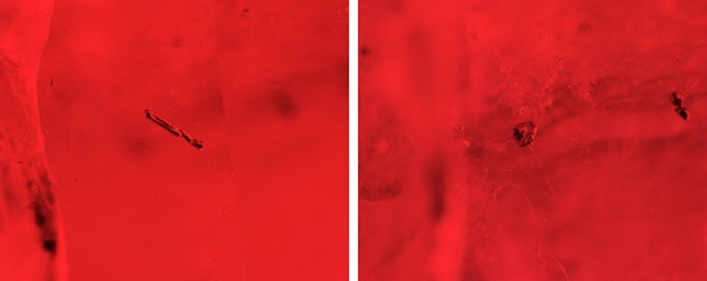
Figure 3. The hexagonal dark reflective long prisms (left) and platelets (right) observed in the sample are common inclusions in flux-grown synthetic corundum. Photomicrographs by Yuxiao Li; field of view 1.28 mm.
Laser ablation–inductively coupled plasma–mass spectrometry (LA-ICP-MS) detected high amounts of Be (35.3–37.1 ppma), Cr (1733.5–2047.2 ppma), Rh (0.6–1.0 ppma), and Pt (0.5–1.1 ppma). Elements normally present in natural corundum such as Mg, Ga, and V were not detected. The chemistry indicated a flux growth process.
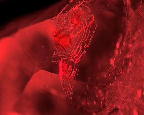
Figure 4. Fingerprints in the flux-grown ruby could also be mistaken for natural residue-filled fingerprints. Photomicrograph by Yuxiao Li; field of view 1.23 mm.
Earlier work has shown numerous transparent, colorless hexagonal tabular “ghost-like” inclusions in Chatham flux-grown sapphire (R.E. Kane, “The gemological properties of Chatham flux-grown synthetic orange sapphire and synthetic blue sapphire,” Fall 1982 G&G, pp. 140–153). In that study, X-ray diffraction analysis (XRD) revealed that the pattern of the ghost-like inclusions was identical to that of chrysoberyl. That study also stated that Chatham, who was successful in synthesizing sapphire by the flux method, added heavy concentrations of beryllium, which explained the presence of chrysoberyl and the high beryllium trace element content. More recently, natural-looking transparent hexagonal crystals in a flux-grown pink sapphire have also been reported (Fall 2017 Lab Notes, pp. 367–368).
There was no surface-reaching transparent crystal in that pink synthetic sapphire, but the small amounts of beryllium suggested the possibility of chrysoberyl. In the present laboratory-grown ruby, fortunately, one of the transparent crystals reached the surface. With Raman spectroscopy, we were able to identify it as chrysoberyl (figure 5).
The beryllium concentration of this laboratory-grown ruby (up to 37 ppma) indicated possible beryllium diffusion treatment. High chromium (up to 2047 ppma) resulted in a vivid red color. With such a highly saturated bodycolor, we cannot deny the possibility of beryllium diffusion, though the signature orange rim was not found using immersion techniques. The numerous chrysoberyl crystals offer important evidence that the beryllium was already excised during the flux growth process. Beryllium diffusion of corundum requires exposure to extreme heat (above 1780°C; J.L. Emmett et al., “Beryllium diffusion of ruby and sapphire,” Summer 2003 G&G, pp. 84–135). These chrysoberyl inclusions do not show the typical indications following heat treatment, meaning this stone was not beryllium diffused after its growth.
With LA-ICP-MS and Raman spectroscopy, we were able to confirm that flux-grown laboratory-grown ruby with high beryllium content could indicate the presence of natural-looking chrysoberyl crystals.